
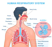
Respiratory System

Presentation
•
Biology
•
Vocational training
•
Practice Problem
•
Hard

Katherine Barajas
FREE Resource
9 Slides • 5 Questions
1
By Ms. B
2

The function of the respiratory system is to move air in and out of the lungs. This process may be called ventilation, respiration, or breathing.
It works with the cardiovascular system to to deliver oxygen to body cells through the bloodstream.
Respiratory System
3
Include:
The nose
pharynx
larynx
trachea
bronchial tree (including bronchi and bronchioles)
lungs
Nostrils are called nares
4

The nasal cavity is the space behind the nose and is divided into left and right portions.
Lined with mucous membrane that warms and moistens air as it passes through the nasal cavity
3 structures called Nasal Conchae extend from the lateral walls and increase the surface area of nasal cavity.
Lined with cilia to move mucus and debris through cavity to pharynx where it is swallowed.
Paranasal sinuses air-filled spaces that reduce weight of the skull and equalize pressure. They also give your voice it's tone.
Nasal Cavity and Paranasal Sinuses (p. 605)
5
Multiple Choice
How many nasal conchae are there?
2
5
3
6
Multiple Choice
The purpose of cilia
Push the mucus and debris toward Pharynx
Tickle your nose
7

Organ of both respiratory and digestive systems.
Consists of 3 separate sections: Nasopharynx (junction of nasal cavity and pharynx), Oropharynx (junction of oral cavity), and Laryngopharynx (contains the larynx or voice box)
During inspiration, air flows from the nasal or oral cavity into the pharynx. From the pharynx, air flows into the larynx.
Pharynx
8
The larynx sits superior to and is continuous with the trachea, or windpipe. It moves air in and out of the trachea and produces the sounds of a person's voice.
Consists mostly of cartilage and muscle tissue
3 Cartilages in the larynx include: Epiglottic cartilage (forms framework of the Epiglottis) The Epiglottis is a flap-like structure that closes off the larynx during swallowing to keep food/liquids do not enter the respiratory system. Thyroid cartilage forms anterior wall of the larynx. Cricoid cartilage forms most of the posterior wall of the larynx.
The vocal cords stretch between the thyroid and cricoid cartilages. The opening between cords is the glottis.
9

The trachea is a tubular organ made of rings of cartilage and smooth muscle. It extends from the larynx to the bronchi.
The distal ends of the trachea branches and starts a series of tubes called the bronchial tree.
The first branches off the trachea are called primary bronchi.
The branches off of this first bronchi branch into tertiary bronchi and then into bronchioles. At the ends of the bronchioles are air sacs called alveoli.
Alveoli are thin sacs made of a single layer of squamous epithelial cells and are surrounded by capillaries. The exchange of oxygen and carbon dioxide takes place.
Through diffusion, red blood cells in the capillaries release carbon dioxide into the alveoli.
Trachea, Bronchi, and Bronchioles
10

The lungs are 2 cone-shaped organs containing connective tissue, the bronchial tree, nerves, lymphatic vessels, and many blood vessels. The right lung is larger than the left because the heart is located in the left thorax.
Right lung divided into 3 lobes (right upper, middle, and lower)
Left lung divided into left upper and lower lobes.
The double-walled membrane that surrounds the lungs is the pleura. The outermost membrane is known as parietal pleura, and innermost is visceral pleura.
The Lungs
11
Breathing consists of inspiration and expiration.
The following events enlarge the thoracic cavity and lead to inspiration: The diaphragm contracts, increasing the space in the thoracic cavity. The intercostal muscles raise the ribs.
During expiration, or exhalation, air rich with carbon dioxide flows out of the airways. When the pressure inside the cavity becomes greater than the atmospheric pressure, aire flows out.
The following events lead to expiration: The diaphragm relaxes. As it does, it domes up into the thoracic cavity, which decreases the space in the cavity. The intercostal muscles lower the ribs, further decreasing the size of the thoracic cavity.
12
Multiple Choice
What does the root Pulmo mean?
Lung
breath
13
Multiple Choice
Trache/o
lung
trachea or windpipe
14
Multiple Choice
plasty
surgical reconstruction
cement
By Ms. B
Show answer
Auto Play
Slide 1 / 14
SLIDE
Similar Resources on Wayground

10 questions
Exercise K-1
Presentation
•
KG
10 questions
Present Perfect Tense
Presentation
•
Professional Development
7 questions
Φθινόπωρο
Presentation
•
KG
9 questions
FIFA Football
Presentation
•
KG
10 questions
W9 Risk 2024
Presentation
•
Professional Development
9 questions
epp lesson
Presentation
•
KG
16 questions
The Theory of Evolution
Presentation
•
8th Grade
12 questions
Review: Incomplete, codomiance and punnet squares
Presentation
•
KG
Popular Resources on Wayground
10 questions
Safety Day 2026 – Student Review

Quiz
•
9th Grade
21 questions
Continents and Oceans
Quiz
•
6th Grade
20 questions
Parts of Speech
Quiz
•
5th Grade
16 questions
Subject & Predicate
Quiz
•
5th Grade
25 questions
Multiplication Facts
Quiz
•
5th Grade
12 questions
Map Skills
Quiz
•
3rd Grade
22 questions
Continents and Oceans
Quiz
•
5th Grade
15 questions
Rounding Decimals
Quiz
•
5th Grade